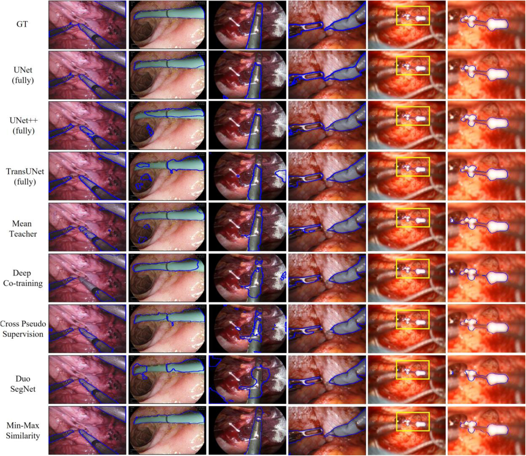
Fig. 2.

Visual comparison of our method with state-of-the-art models. Segmentation results are shown for 50% of labeled training data for Kvasir-instrument, EndVis’17, ART-NET and RoboTool, and 2.4% labeled training data for cochlear implant. From left to right are EndoVis’17, Kvasir-instrument, ART-NET, RoboTool, Cochlear implant and region of interest (ROI) of Cochlear implant.
