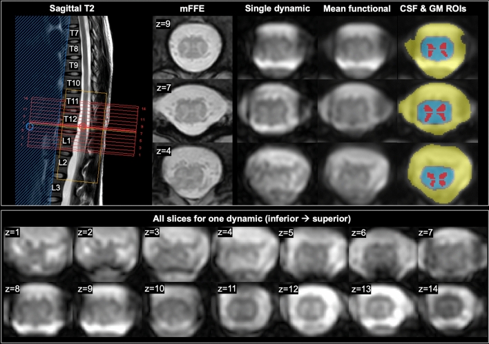
Figure 1.
Example acquired images in one participant (26-year-old female). Left: acquired volume (red box) centered at the lumbar enlargement, shown on a sagittal T2-weighted scan. A multi-echo gradient echo scan (proton-density/T2*-weighted axial multi-echo fast field echo (mFFE), acquired voxel size 0.65 × 0.65 × 5mm3) is included for visualization of anatomy. Middle to right: a single functional dynamic after motion correction; the mean functional image after denoising; and regions of interest including cerebrospinal fluid (CSF) in yellow (used for nuisance regression), cord in blue, and gray matter (GM) horns in red. Representative slices for one dynamic volume in one participant are shown below, and numbered (z) from inferior (caudal) to superior (rostral).

