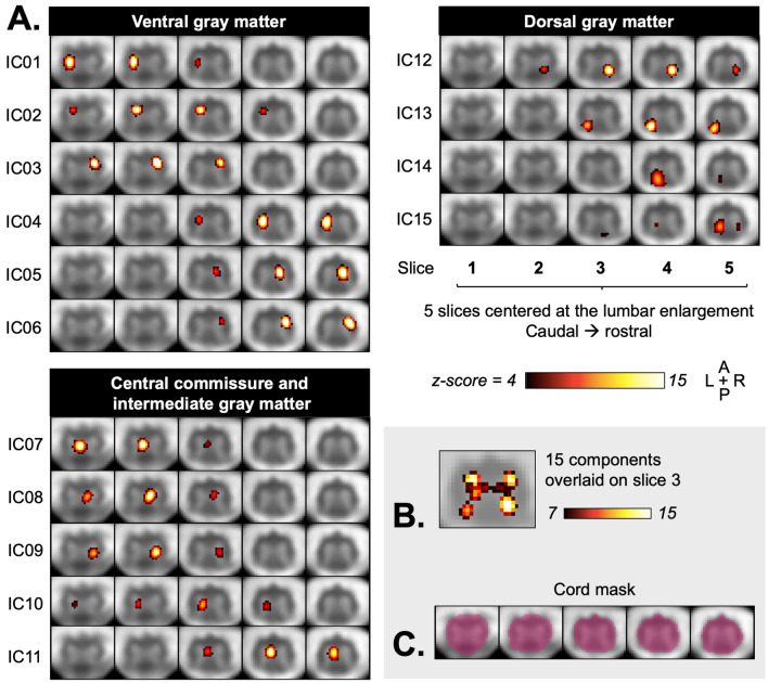
Figure 5.
Group spatial independent component analysis (ICA)-derived components from 26 participants. (A) Aggregate group component maps are shown, overlaid on the group-average functional image, with each row showing one independent component across five slices (IC). Six ventral, 4 dorsal, and 5 central commissure/intermediate gray matter components are shown, organized top-to-bottom by their location from rostral to caudal and left to right. (B) Overlaid components for the middle slice (slice 3), thresholded at z ≥ 7, show the shape of the gray matter. (C) The cord mask used for analysis is shown in pink.

