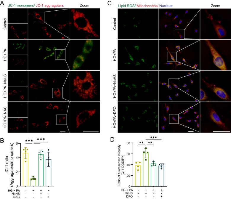
Fig. 4. Exogenous H2S protects cardiac mitochondria.
A Mitochondrial membrane potential (MMP) of HL-1 cardiomyocytes detected by fluorescence probe JC-1. Scale bar: 50 μm. B Quantification of JC-1, n = 4. C Lipid ROS levels and locations in HL-1 cardiomyocytes detected by C11-BODIPY. Scale bar: 50 μm. D Quantification of lipid ROS, n = 4. All quantitative data are presented as mean ± SD from independent experiments. *P < 0.05, **P < 0.01, ***P < 0.001 by ordinary one-way ANOVA.

