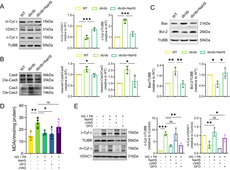
Fig. 5. Exogenous H2S prevents cardiomyocytes from mitochondrial apoptosis.
A WB showing mitochondrial cytochrome c (m-Cyt c) and cytoplasmic cytochrome c (c-Cyt c) levels in cardiac tissues, n = 3. B The protein levels of Bax and Bcl-2 in cardiac tissues, n = 3. C Cleaved-Caspase-9 (Cle-Cas9) and cleaved-Caspase-3 (Cle-Cas3) in cardiac tissues, n = 3. D MDA concentration in HL-1 cardiomyocytes, n = 4. E WB showing c-Cyt c and m-Cyt c levels of HL-1 cardiomyocytes, n = 3. All quantitative data are presented as mean ± SD from independent experiments. *P < 0.05, **P < 0.01, ***P < 0.001 by ordinary one-way ANOVA.

