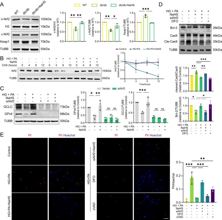
Fig. 6. Exogenous H2S inhibits apoptosis through Nrf2.
A WB showing nuclear Nrf2 (n-Nrf2), cytoplasmic Nrf2 (c-Nrf2) and Keap1 levels in cardiac tissues, n = 3. B Degradation of Nrf2 in HL-1 cardiomyocytes. Cells were treated with cycloheximide (CHX, 100 µM) at 0, 4, 8 and 12 h after other treatment, n = 3. C Protein levels of GCLC and GPx4 in HL-1 cardiomyocytes, n = 3. D The protein levels of Bcl-2 and cleavage of Cas9 in HL-1 cardiomyocytes, n = 3. E Cell death determined by PI/Hoechst staining in cardiomyocytes, n = 3. Scale bar: 100 μm. All quantitative data are presented as mean ± SD from independent experiments. *P < 0.05, **P < 0.01, ***P < 0.001 by ordinary one-way ANOVA.

