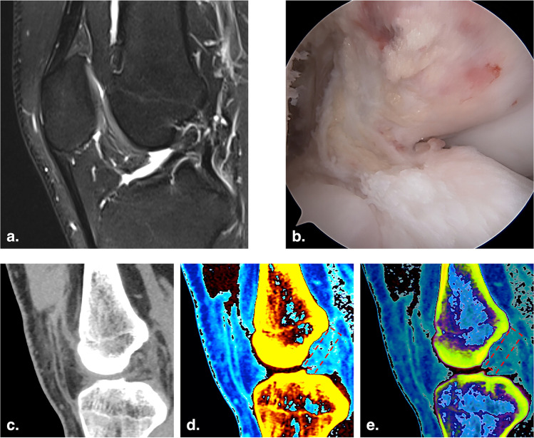
Fig. 2.
Images of a 22-year-old man with right ACL rupture. a Sagittal T2-weighted fat-saturated MRI image of the right knee. b Arthroscopic image of the torn right ACL. c Monochromatic DECT image with normal mode (mixed keV) on the oblique sagittal plane. d and e Color-coded DECT images with mono + mode (80 keV) and Rho/Z mode (mixed keV) on the oblique sagittal plane, respectively. The torn ACL was not colored in the anatomic position (red dotted rectangle)

