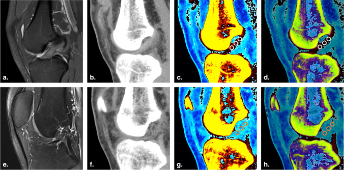
Fig. 3.
Images of a 25-year-old man with left ACL rupture. a and e Sagittal proton density-weighted and T2-weighted fat-saturated MRI images of the right and left knees, respectively. b and f Oblique sagittal plane, monochromatic DECT images with normal mode (mixed keV) of the right and left knees, respectively. c and g Oblique sagittal plane, color-coded DECT images with mono + mode (80 keV) of the right and left knees, respectively. d and h Oblique sagittal plane, color-coded DECT image with Rho/Z mode (mixed keV) of the right and left knees, respectively. The intact ACL was colored black and dark red in the anatomic position (c and d). The torn ACL was not colored (g and h). Three circular ROIs were set on the proximal, middle, and distal sites of the normal ACL (c and d) and torn ACL (g and h), respectively

