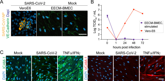
Fig. 3.
Inflammatory conditions do not alter interaction of SARS-CoV-2 with EECM-BMECs. (A) Representative confocal images from immunofluorescence staining for SARS-CoV-2 NP (orange) and VE-cadherin (green) 72 hpi with SARS-CoV-2 at 50’000 TCID50/ well or Mock in EECM-BMECs, which were pre-stimulated with TNFα (1 ng/mL) and IFNγ (20 IU/mL) for 20 h, are shown. Nuclei were stained with DAPI (blue). Three iPSC-clone-derived EECM-BMECs with 2 replicates per condition were tested. As a positive control VeroE6 cells 72 hpi with SARS-CoV-2 at 400 TCID50/ well is shown. Scale bar = 50 μm. (B) Quantification of released virions into the supernatant 1–72 hpi with SARS-CoV-2 by TCID50 assay. Measurement was done in duplicates from a total of 3 experiments with EECM-BMECs (each time a different iPSC clone) and 1 experiment with VeroE6 cells. (C) Representative confocal images of immunofluorescence staining for cell adhesion molecules VCAM-1 (red) and ICAM-1 (green) in EECM-BMECs after 24 h inoculation with Mock, SARS-CoV-2 or as a positive control stimulation with TNFα (1 ng/mL) and IFNγ (20 IU/mL) are shown. Nuclei were stained with DAPI (blue). Scale bar = 50 μm. 3 iPSC clone-derived EECM-BMECs with 2 replicates per condition were tested

