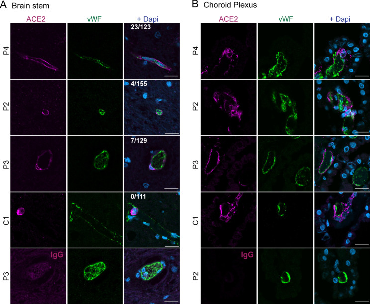
Fig. 5.
ACE2+ cells localized next to endothelial cells in brain stem and ChP in COVID-19 patients. A-B) Representative confocal images from immunofluorescence staining of ACE2 (magenta) and von Willebrand factor (vWF, green) of brain stem (medulla oblongata) (A) and choroid plexus (B) from 3 COVID-19 patients and 1 control. Nuclei were stained with DAPI (blue). Scale bar = 20 μm. The numbers indicated in the merged image of A indicate the number of vWF+ vessels with ACE2 signal / total number of vWF+ vessels. Quantification was done from 15 images per patient

