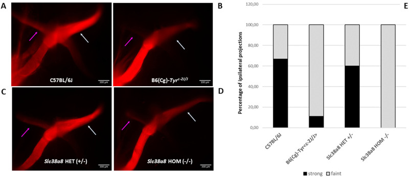
Figure 6.
Analysis of retinal ganglion cell projections of Slc38a8 mutant mice. Unilateral (left eye) anterograde labeling of +18.5 dpc embryos with neuronal tracer DiI. (A) Representative images of whole-mounted optic chiasms from wild-type pigmented C57BL/6J, albino B6(Cg)-Tyrc-2J/J, Slc38a8 heterozygous “HET” (+/–), and Slc38a8 homozygous mutant “HOM” (–/–) mice. Ipsilateral projections are indicated with a purple arrow. Contralateral projections are indicated with a white arrow. (B) Quantification of ipsilateral projections. Bars are expressed in percentages considering less intense stained (barely visible, shown in grey) or more intense stained (clearly visible, shown in black) uncrossed fibers. Data are derived from N = 12 (pigmented), N = 11 (albino), N = 12 (Slc38a8 +/–), and N = 9 (Slc38a8 –/–) number of fetuses analyzed. Ipsilateral projections were detected only in N = 9 (pigmented, 6 strong and 3 faint), N = 9 (albino, 1 strong and 8 faint), N = 10 (Slc38a8 +/–, 6 strong and 4 faint), and N = 4 (Slc38a8 –/–, 0 strong and 4 faint). Fluorescent pictures are presented unaltered, as provided by the standard automated capture system (computer program LasX, Leica), with identical parameters for all individuals.

