Figure 9.
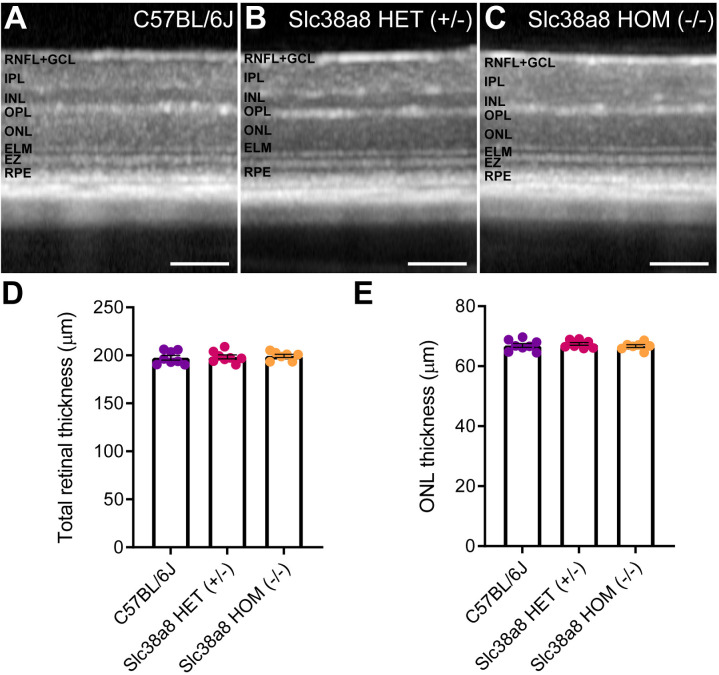
Retinal structure of Slc38a8 mutant mice evaluated in vivo by optical coherence tomography. Representative OCT images from control pigmented C57BL/6J (A), heterozygous Slc38a8 (+/–), (B) and homozygous Slc38a8 (–/–), (C) mutant mice. (D) Quantification of the total retinal thickness. (E) Quantification of the ONL thickness (outer nuclear layer, photoreceptor nuclei). Mean ± SEM, N = 5 to 8. Two-way ANOVA with Bonferroni correction. There are no statistically significant differences. RNFL, retinal nerve fiber layer; GCL, ganglion cell layer; IPL, inner plexiform layer; INL, inner nuclear layer; OPL, outer plexiform layer; ELM, external limiting membrane; EZ, ellipsoids zone; RPE, retinal pigment epithelium. Scale 100 µm.

