Figure 1.

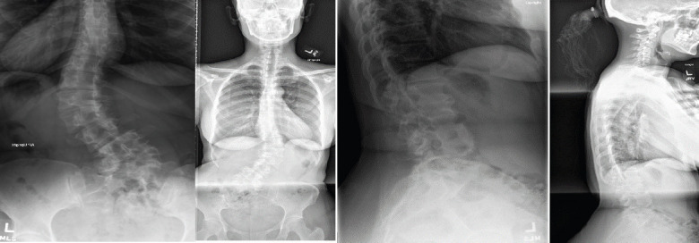
Pre-operative coronal (left) and lateral (right) radiographs. Radiographs demonstrate a 50° thoracolumbar curve from T11-L5, a right L5 hemivertebrae, lateral listhesis at L3-4, 29° of lumbar lordosis and truncal shift to the left.

Pre-operative coronal (left) and lateral (right) radiographs. Radiographs demonstrate a 50° thoracolumbar curve from T11-L5, a right L5 hemivertebrae, lateral listhesis at L3-4, 29° of lumbar lordosis and truncal shift to the left.