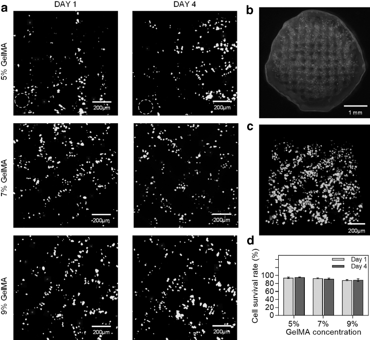
FIG. 5.
The effect of GelMA concentration on the cell survival rate. (a) Live/Dead staining of GelMA printing with a concentration of 5%, 7%, and 9% (living cells were depicted in green and dead cells in red, 10 × ). (b) Overall diagram of MNGCs with the concentration of 7%. (c) The 3D local view of MNGCs. (d) The quantification of the survival rate of SCs. SCs, Schwann cells.

