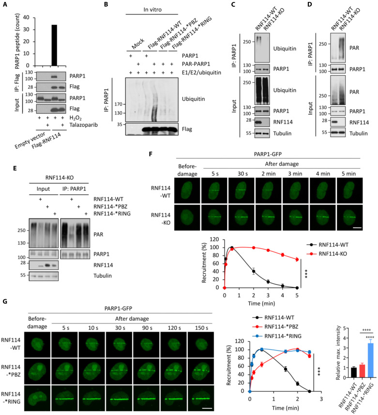
Fig. 3. RNF114 targets PARylated-PARP1 for ubiquitin-proteasomal degradation.
(A) RNF114 interacts with PARylated-PARP1. HCT116 cells expressing the empty vector or Flag-RNF114 were pretreated with talazoparib (1 μM for 1 hour) and were treated with H2O2 (2 mM for 5 min). The whole-cell lysates were subjected to immunoprecipitation (IP) (anti-Flag), and the immunoprecipitants were analyzed by liquid chromatography tandem mass spectrometry (LC-MS/MS) experiments (top) and immunoblot experiments (bottom). (B) In vitro ubiquitination assays of PARP1 or PARylated-PARP1. Purified RNF114-WT, RNF114-*PBZ mutant, or RNF114-*RING mutant was subjected to in vitro ubiquitination experiments in the presence of PARylated-PARP1. (C) RNF114 mediates the ubiquitination of PARP1. RNF114-WT and RNF114-KO HCT116 cells were pretreated with MG132 (10 μM for 6 hours) and then were treated with H2O2 (2 mM for 5 min). PARP1 was isolated using IP and was subject to immunoblotting analyses. (D) RNF114 mediates the degradation of PARylated-PARP1. RNF114-WT and RNF114-KO HCT116 cells were treated with H2O2 (2 mM for 5 min). PARP1 was isolated using IP, and was subject to immunoblotting analyses. (E) RNF114 with the uncompromised PAR-binding and E3 ligase activity is required for the degradation of PARylated-PARP1. RNF114-KO HCT116 cells were reconstituted with RNF114-WT, RNF114-*PBZ mutant, or RNF114-*RING mutant. These cells were pretreated with H2O2 (2 mM for 5 min). PARP1 was isolated using IP and was subjected to immunoblotting analyses. (F) Deletion of RNF114 leads to PARP1 trapping. RNF114-WT and RNF114-KO HeLa cells expressing green fluorescent protein (GFP)–tagged PARP1 were subjected to the laser microirradiation experiment. (G) Interference with the PAR-binding or the E3 activity of RNF114 leads to PARP1 trapping. RNF114-KO HeLa cells were reconstituted with the RNF114-WT, RNF114-*PBZ mutant, or RNF114-*RING mutant. These cells were transfected with GFP-tagged PARP1 and were subjected to a laser microirradiation assay. GFP signals were monitored and were quantified in a time-course experiment. Scale bars, 10 μm. KO, knockout.

