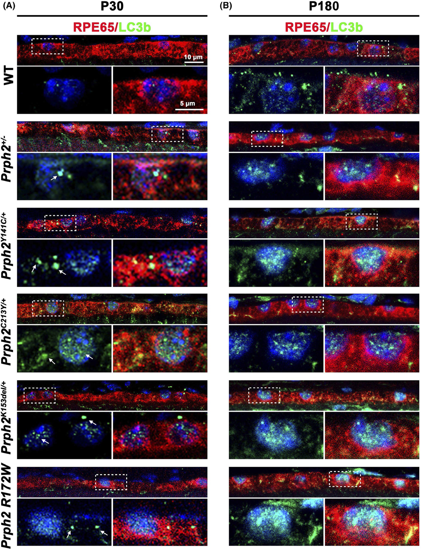
FIGURE 5.

LC3b positive aggregates increase in Prph2 disease models. (A, B) Retinal sections harvested at P30 (A) and P180 (B) were co-labeled with LC3b (green) and RPE65 (red) and counterstained with DAPI (blue). Dotted outlined areas are magnified in panels shown below. Arrows highlight accumulation of LC3b aggregates in (A). Magnification 63×. Scale bars: 10 μm (top images) and 5 μm (insets) N = 3 eyes per genotype and age
