
FIGURE 2.
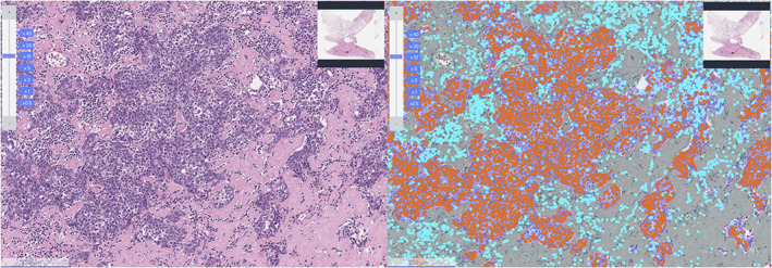
Representative image of Lunit SCOPE IO. Representative image of hematoxylin and eosin (H&E) original image (left) and Lunit SCOPE IO‐inferenced segmentation of cancer epithelium (orange), cancer stroma (grass green), and TIL (cyan blue), respectively (right).