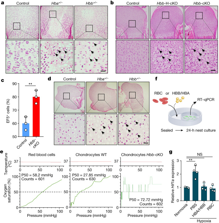
Fig. 4. Haemoglobin is essential for chondrocyte hypoxia adaption and survival of the fetal cartilage.
a, Histological examination of proximal humeral cartilages from P5 mice of different genotypes. The arrows indicate dead chondrocytes. Scale bar, 50 μm. n = 6 biologically independent samples. b, Histological images of proximal humerus from newborn mice upon cKO of the Hbb gene. Control: Hbb+/+/Col2a1-CreERT2 mice, Hbb-H-cKO: HbbF/+/Col2a1-CreERT2 mice (heterozygous deletion), and Hbb-cKO: HbbF/F/Col2a1-CreERT2 mice. The arrows indicate dead chondrocytes. Scale bar, 50 μm. n = 6 biologically independent samples. c, Quantification of EF5-positive cells in cartilages from WT and Hbb-cKO mice. n = 3 biologically independent samples for each. Error bars represent s.e.m. **P = 0.0080. d, Histological examination of cartilages of E18.5 mice cultured in vitro under hypoxia (1% O2) for 3 days. Black arrows indicate the dead chondrocytes. Scale bar, 50 μm. e, Oxygen dissociation curves of RBCs, WT and Hbb-cKO chondrocytes. The chondrocytes of Hbb-cKO displayed poor oxygen-binding capability as indicated by the fluctuated curve. Counts indicate the measurement time (in seconds), the horizontal grey lines indicate oxygen partial pressures in the environment of chondrocytes. f, Schematic diagram of the nest co-culture experiment, in which the hypoxia-responsive cells were cultured in the inner dish, whereas the RBCs or the haemoglobin condensates were placed in the outer dish that was sealed for 24 h to create hypoxic conditions. g, Expression of Hif1a under the indicated conditions of nest co-culture as examined by qPCR. The data are mean ± s.e.m. of triplicate experiments; two-sided Student’s t-test was used for the data analysis, and adjustment was not made for multiple comparisons. **P = 0.0019 (phosphate-buffered saline), P = 0.6454 (HBA/HBB) and P = 0.4890 (RBC). NS, not significant.

