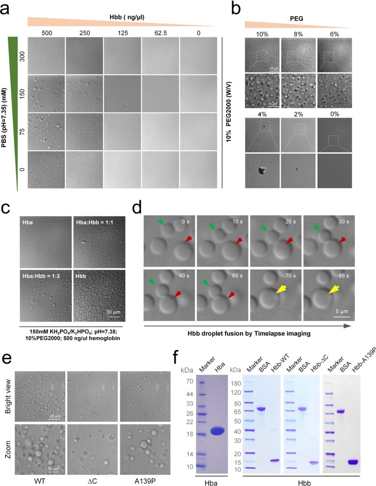
Extended Data Fig. 3. Liquid phase separation of the purified untagged Hbb in vitro.
a, Droplet formation of the purified untagged Hbb in different conditions, buffer (PBS: KH2PO4/K2HPO4, pH 7.35, PEG2000 10% (w/v)). Scale bar: 20 μm. b, Droplet formation of the purified untagged Hbb at different PEG concentrations in phase separation buffer (150 Mm KH2PO4/K2HPO4, pH 7.35, PEG2000 variable (w/v)). Scale bar: 20 μm. c, Droplet formation of the purified untagged Hba, Hbb and the mixture of them at the same protein concentration in phase separation buffer (150 mM KH2PO4/K2HPO4, pH 7.35, PEG2000 10% (w/v)). Scale bar: 30 μm. d, Timelapse imaging of the purified untagged Hbb droplet fusion. Scale bar: 5 μm. e, Droplet formation of the purified Hbb and its IDR mutants in phase separation buffer (150 mM KH2PO4/K2HPO4, pH 7.35, PEG2000 10% (w/v)). Scale bar: 25 μm, 10 μm. f, Coomassie brilliant blue stained gels of the purified Hba, Hbb and Hbb mutants.

