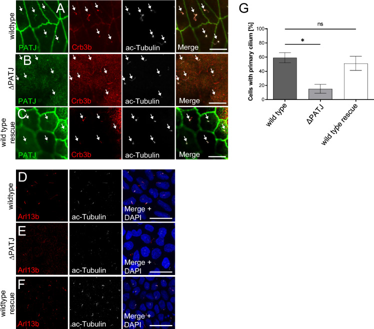
Fig. 2.
PATJ localizes to primary cilia and regulates cilia maintenance. A–C Immunostainings of PATJ, acetylated α-Tubulin (Ac-Tubulin) and Crb3b in wild-type MDCK cells (A), PATJ-deficient cells (B) and MDCK∆PATJ-cells with mPATJ-GFP rescue (C). Arrows indicate primary cilia. D–F Staining of primary cilia with Arl13b and ac-Tubulin demonstrates a reduction in primary cilia. G Quantification of primary cilia in the indicated cell lines. Error bars are standard errors of the means. Significance was determined by one-way ANOVA test and Bonferroni correction: *p < 0.05, ns not significant. Scale bars are 10 µm in A–C and 20 µm in D–F

