Fig. 6.
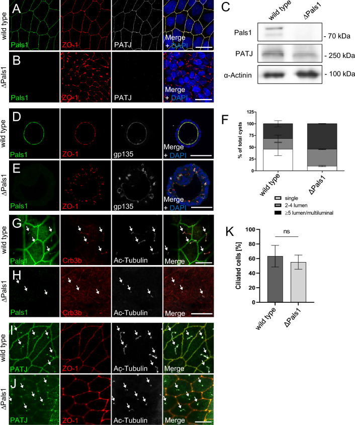
Loss of Pals1 results in TJ and lumen formation defects but does not affect cilia formation. A, B Immunostaining of wild-type MDCK cells (A) and Pals1-deficient MDCK cells (B) with the indicated antibodies. C Western blot analysis of wild-type MDCK and MDCK∆Pals1 cells. D, E Immunostaining of wild-type MDCK (D) and MDCK∆Pals1 (E) cells cultured in Matrigel with the indicated antibodies. F Quantification of lumen formation in the indicated cell lines. Error bars are standard errors of the means. G Pals1 colocalizes with Crb3b at primary cilia, which are labelled with ac-Tubulin. H Immunostaining of MDCK∆Pals1 cells with Pals1, Crb3b and ac-Tubulin. I, J PATJ is mostly lost from TJ in Pals1-depleted cells (J) but still localizes to primary cilia (arrows). L Quantification of primary cilia number in wild-type and Pals1-deficient MDCK cells reveals no significant differences. Error bars are standard errors of the means. Scales bars are 20 µm in A–B and G–J and 50 µm in D–E

