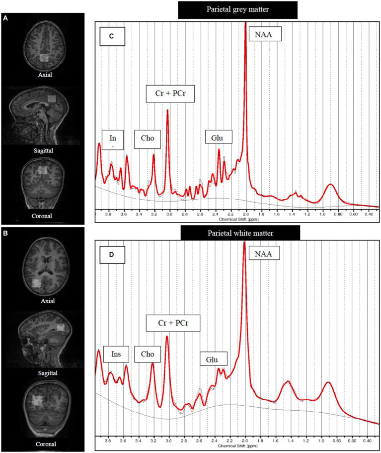
Figure 1.
Structural MRI images showing MRS voxel placements and example SVS spectra [LCModel output]. Axial, sagittal and coronal views of the single voxel (25 × 25 × 25 mm3) placed covering (A) parietal gray matter and (B) right hemisphere parietal white matter. Examples of (C) parietal gray matter and (D) parietal white matter MRS spectra. NAA, N-acetyl aspartate; Glu, Glutamate; GPC+PCh, total choline (glycerophosphocholine +phosphocholine); Cr+PCr, total creatine (creatine +phosphocreatine).

