
Figure 1.
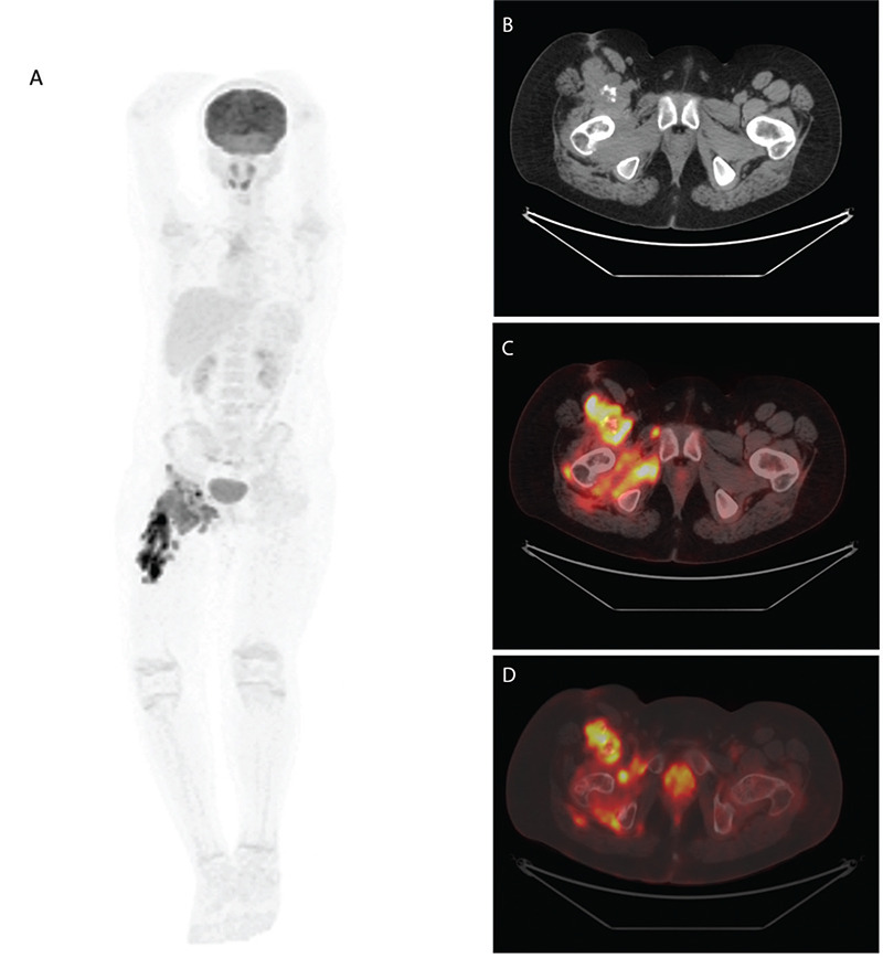
A 13-year-old male patient with an inflammatory myofibroblastic tumor of the right thigh. Maximum intensity projection 18F-fluorodeoxyglucose (18F-FDG) positron emission tomography (PET) image (A), axial computed tomography (CT) (B), and axially fused 18F-FDG PET image (C) showed increased 18F-FDG uptake [maximum standardized uptake value (SUVmax): 28.60] in the multifocal mass with heterogeneous calcifications through the anterior part of the femur. Bone structure findings in the axial CT bone window (D) are within normal limits.