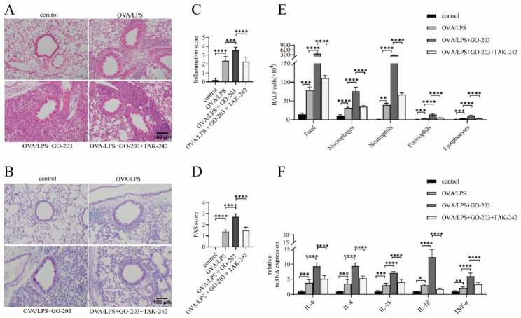
Fig. 6.
TAK-242 partially ameliorates GO-203-exacerbated neutrophilic airway inflammation in OVA/LPS-induced asthmatic mouse. (A, B) HE staining and PAS staining were performed on lung sections in the indicated groups. Images were taken under a microscope at 200× magnification. Scale bar: 50 μm. (C, D) Inflammatory infiltration and hyperplasia of goblet cells were quantified using inflammation and PAS scores. (E) Statistical analysis of the total inflammatory cells, macrophages, neutrophils, eosinophils, and lymphocytes in BALF. (F) The mRNA expression levels of IL-6, IL-8, IL-18, IL-1β, and TNF-α in lung tissues were detected using RT-qPCR. LDH = lactate dehydrogenase. Data are expressed as mean ± SD (n = 6 animals per group) and were analyzed using one-way analysis of variance,*p < 0.05; **p < 0.01; ***p < 0.001; and ****p < 0.0001

