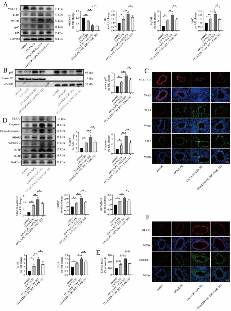
Fig. 7.
TAK-242 downregulates GO-203-caused TLR4/MyD88/NF-κB pathway activation and NLRP3 inflammasome-mediated pyroptosis in OVA/LPS-induced asthmatic mouse. (A) The protein expression levels of MUC1, TLR4, MyD88, p-p65, and p65 in lung tissue were determined via immunoblotting. (B) The expression of MUC1, TLR4, and p-p65 in the bronchi were determined via immunofluorescence staining. Images were taken using a fluorescent microscope at 400× magnification. Red: MUC1; green: TLR4 and p-p65; blue: DAPI; scale bar: 50 μm. (C) The p65 protein levels in the nuclear and cytoplasmic fractions in lung tissue were analyzed via immunoblotting. (D) The protein expression levels of NLRP3, caspase-1, cleaved caspase-1, GSDMD, GSDMD-N, IL-18, and IL-1β in lung tissue were determined via immunoblotting. (E) LDH release into BALF was detected via LDH release assay. (F) The expression of NLRP3 and caspase-1 in the bronchi were determined via immunofluorescence staining. Images were taken using a fluorescent microscope at 400× magnification. Red: NLRP3; green: caspase-1; blue: DAPI; scale bar: 50 μm. LDH = lactate dehydrogenase. Data are expressed as mean ± SD (n = 6 animals per group) and were analyzed using one-way analysis of variance, *p < 0.05; **p < 0.01; ***p < 0.001; and ****p < 0.0001

