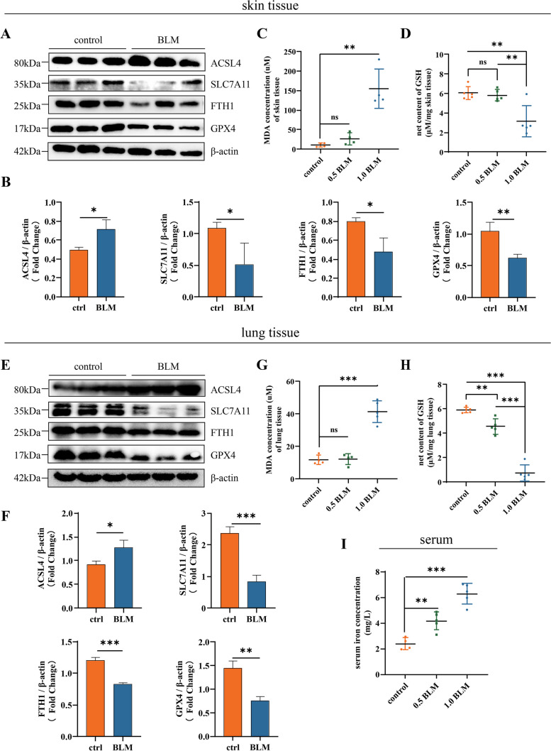
Fig. 1.
Ferroptosis is present in the BLM-induced SSc mice model. A, B Western blots for ferroptosis marker of the skin tissue from healthy control mice and BLM-induced mice (1 mg/ml, 0.1 ml/day, for 4 weeks). C Relative MDA levels of murine skin tissue in healthy control group, moderate BLM group (0.5 mg/ml BLM, 0.1 ml/day, for 4 weeks), and severe BLM group (1 mg/ml BLM, 0.1 ml/day, for 4 weeks) (n = 5). D Net GSH concentration of murine skin tissue in above groups (n = 5). E, F Western blots for ferroptosis marker of the lower back lung tissue from healthy control mice and BLM-induced mice (1 mg/ml, 0.1 ml/day, for 4 weeks). G Relative MDA levels of murine lung tissue in healthy control group, moderate BLM group, and severe BLM group (n = 5). H Net GSH concentration of murine lung tissue in above groups (n = 5). I Serum iron level of each group (n = 5). (Data are presented as Mean ± SD *p < 0.05, **p < 0.01, ***p < 0.001)

