Fig 3.
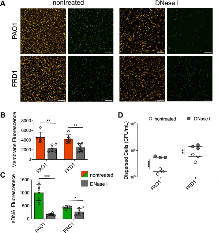
DNaseI disrupts mucoid biofilms and disperses bacterial cells. Representative images of bacterial membrane and eDNA-stained nonmucoid PAO1 and clinical mucoid FRD1 biofilms, nontreated (left) or treated (right) with DNaseI (200 µg/mL) after 16-h biofilm formation. The left panel depicts merged images of FM4-64 and TOTO-1 channels, the right panel depicts the TOTO-1 channel alone; the scale bar is 20 µm (A). Quantification of FM4-64 (bacterial membrane; orange) (B) and TOTO-1 (eDNA; green) (C) fluorescence using NIS-elements image analysis software comparing DNaseI treated (gray) counterparts for each strain. CFU quantification of bacterial cells from supernatant of biofilms treated with or without DNaseI (D). N = 6, Analyzed using student t-test *P < 0.05, **P < 0.005, ***P < 0.0005.

