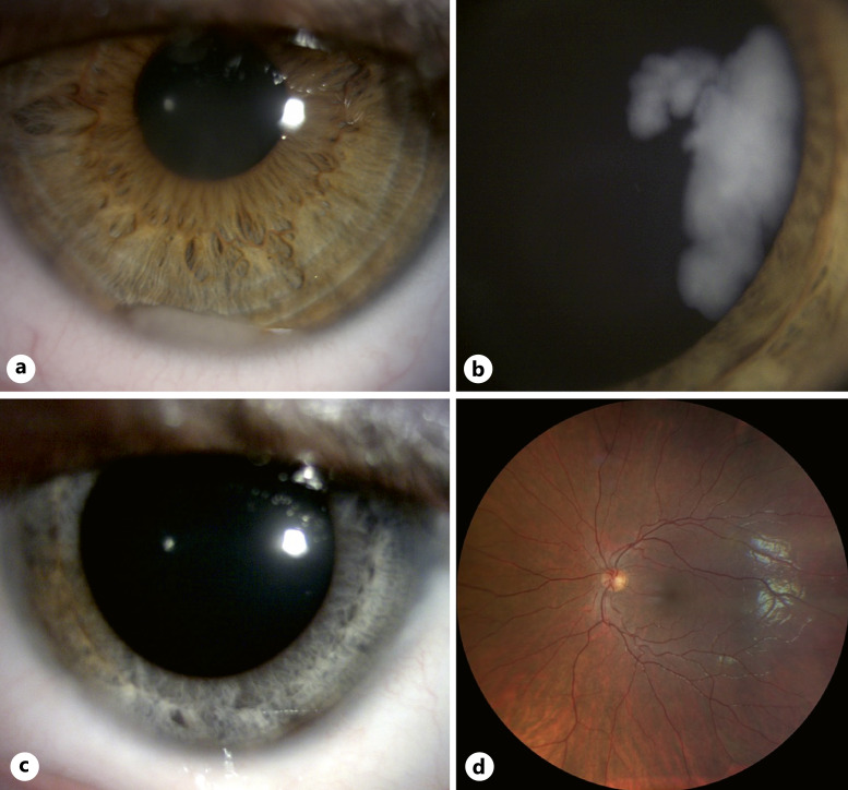
Fig. 1.
DIR with anterior involvement in an 8-year-old boy. a Anterior segment at diagnosis showing an inferior cellular pseudo-hypopyon. b Wide-field fundus color photograph at diagnosis showing diffuse tumoral infiltration of the peripheral retina and ciliary body. c Anterior segment photograph taken 2 years after the last treatment received showing diffuse iris discoloration. d Normal wide-field fundus photograph 2 years after the last treatment received.

