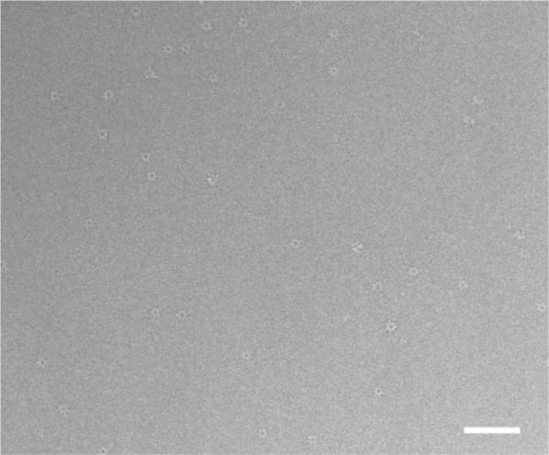
Figure 1. Cryo-EM Images Used in Reconstruction.

A micrograph showing a field of the PA63h.LF complex suspended in vitrified buffer over a hole in a carbon film and recorded at nominal defocus of 2.0 μm. The protein is displayed with a lighter density compared to the surrounding vitrified buffer, i.e., in reversed contrast. Both en face views and relatively fewer “side views” can be seen. Excess (unbound) LF molecules contribute to the background. (Bar = 1000 Å ).
