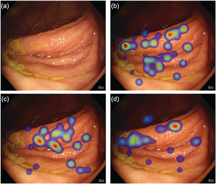
Figure 2.

Eye‐tracking outputs for different participants viewing the same image. (a) Raw image with sessile serrated lesion highlighted in subsequent images with blue bounding box. (b) Correct detection with black cross indicating that the participant marked the polyp manually, corroborated with eye‐tracking heat map demonstrating fixations in the polyp region defined by the blue bounding box. The heat map colors represent the cumulative number of fixations, with red indicating a longer gaze (more fixations). (c) A gaze error is demonstrated; the participant did not mark the polyp manually (absence of cross) and also did not fixate on the region; that is, polyp was not observed. (d) A cognitive error is demonstrated; the participant did not mark the polyp manually (absence of cross) but fixated in the polyp region defined by the blue bounding box, suggesting that the polyp was observed.
