
Figure 5 |. Immunofluorescence staining showing localization of ENPP1, IGFBP3, ARID5B and ADK in control and atherosclerotic human coronary arteries.
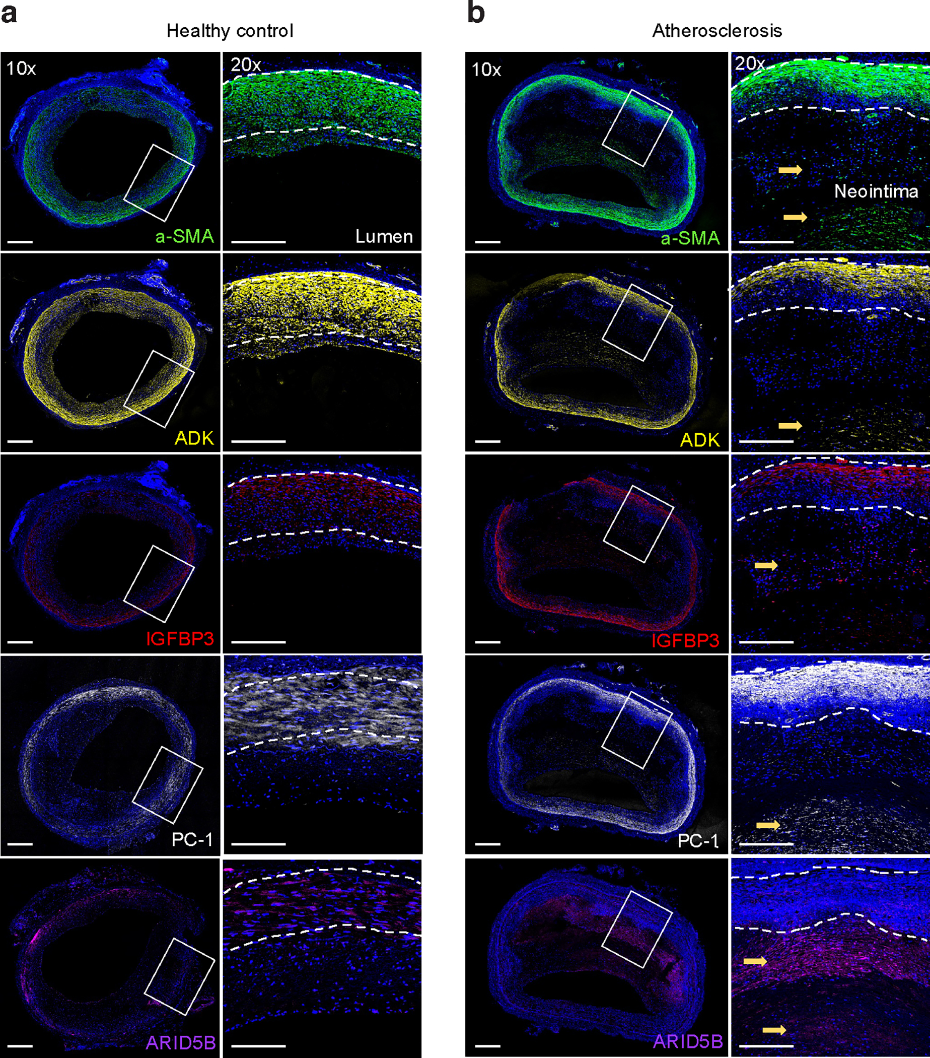
a,b, Transverse sections of healthy control (a) and atherosclerotic (b) human coronary arteries were stained for alpha-smooth muscle actin (α-SMA) (green), DAPI nuclei marker (blue), ENPP1/PC-1 (white), IGFBP3 (red), ARID5B (purple), and ADK (yellow). High levels of ENPP1/PC-1, IGFBP3, ARID5B, and ADK were observed in the neointimal layer of atherosclerotic diseased coronary arteries. Whole artery images were captured at 10× magnification and regions of interest were captured at 20×. Images are representative of n = 4 independent donors per group.