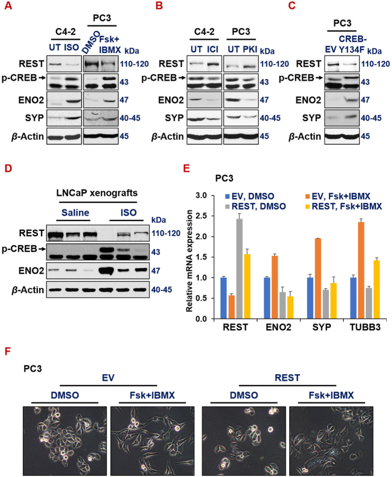
Figure 4. Activated CREB signaling represses REST.
(A-B) Western blotting for REST, p-CREB1 (pS133, indicator of activation) and NE markers in prostate cancer cells (C4-2 on left and PC3 on right) upon treatments of CREB1 signaling activator 15uM isoproterenol (ISO) or 10uM Forskolin+ 0.5mM IBMX for 24hr (A), or CREB1 signaling inhibitor ICI-118,551 (ICI, 10uM) or synthetic peptide inhibitor of PKA (PKI, 10uM) for 24hr (B). (C) Western blotting for REST, pS133-CREB1 and NE markers in PC3 cells carrying either an empty vector or CREB1-Y134F cDNA, a constitutively activated form of CREB1. (D)Western blotting for REST, pS133-CREB1, NE markers, EZH2 catalytic product H3K27me3 histone mark, loading controls histone 3 (H3) and beta actin in LNCaP cell-derived xenografts from NOD/SCID male mice treated with saline or 10mg/kg ISO for 54 days. (E-F) PC3-EV or PC3-REST cells were treated with DMSO control or CREB1 signaling activator combo 10uM Forskolin (Fsk) + 0.5mM IBMX for 24hr. mRNA levels of indicted genes were normalized to GAPDH controls. (F) Morphology of PC3-EV and PC3-REST cells treated with DMSO or Fsk+IBMX.

