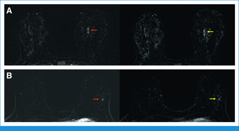
FIG 3.
The HTHS protocol facilitates cancer detection in patients with marked BPE. (A) Forty-three-year-old woman with a left breast focal nonmass enhancement in the central left breast. The nonmass enhancement is well appreciated on a T1-weighted fat-saturated subtraction image the third postcontrast phase of the HTHS protocol (red arrow), acquired 36 seconds after contrast injection. The lesion becomes obscured by marked BPE on the seventh postcontrast phase of the HTHS protocol (yellow arrow), acquired 84 seconds after contrast injection. Pathology yielded HR+/HER2– high-grade invasive lobular carcinoma. (B) Forty-nine-year-old women with focal nonmass enhancement in the outer left breast, which is well appreciated on a T1-weighted fat saturated subtraction image during the third postcontrast phase of the HTHS protocol (red arrow), acquired 36 seconds after contrast injection. The lesion is obscured by marked BPE on the seventh postcontrast phase of the HTHS protocol (yellow arrow), acquired 84 seconds after contrast injection. Pathology yielded HR+ high-grade ductal carcinoma in situ. BPE, background parenchymal enhancement; HER2, human epidermal growth factor receptor 2; HR, hormone receptor; HTHS, high temporal/high spatial resolution.

