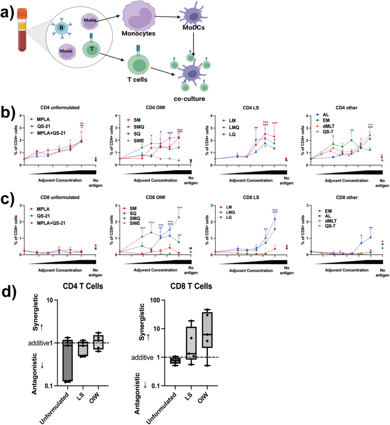
Fig. 4. MoDCs stimulated with co-formulated MPL and QS-21 activate SARS-CoV-2 Spike protein-specific T cells in DTI assay.
a Schematic representation of the DC:T cell interface (DTI) assay. b, c MoDCs were stimulated with 5 μg/mL SARS-CoV-2 Spike antigen, in the presence or absence of adjuvant formulations at a 1:100 dilution for LNP, OIW, and Other, and 1:1000 dilution for Soluble formulations, and subsequent 1/6 (v/v) dilutions for all formulations) before co-culture with autologous CD4+ or CD8+ T cells. Activated T cells were defined as CD4+, CD8+, CD25+, CD134+, or CD154+. Mean + SD are plotted for each concentration. d Calculated D-values quantify the extent of adjuvant interaction (i.e., antagonism, additivity, or synergy) between MPL and QS-21 in each formulation type. (Box-whisker plots: centre line: median; bounds: 25th/75th percentile; whiskers; lowest/highest point) Three data points were excluded (2 soluble, 1 OIW) because a curve could not be fit using the method described. Stars above a data point on dose-response curves indicate statistical significance relative to vehicle control, stars adjacent to vertical bars on dose-response curves indicate statistically significant differences between treatment groups at a given concentration, and stars above horizontal lines indicate statistically significant difference relative to a control with no antigen. Significance was calculated using a two-way repeated measures ANOVA with Tukey’s multiple comparison testing. N = 5 for all assays. (*p < 0.05, **p < 0.01, ***p < 0.001).

