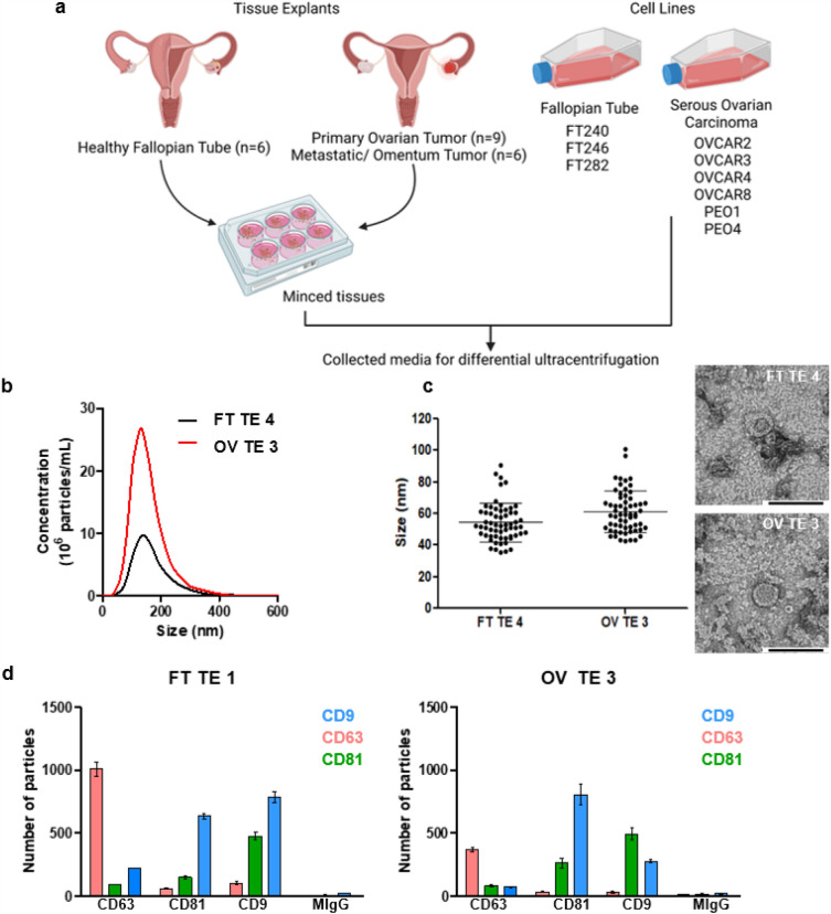
Figure 1.
Enrichment and characterization of cell line and tissue explant EVs. (a) Surgical resections of healthy FT or tumor tissues were minced and used to initiate short-term tissue explants (cultured for 24 h) followed by collection of the conditioned media and processed by differential ultracentrifugation to enrich for EVs. Likewise, conditioned media from the FT and ovarian cancer cell lines shown was collected and processed. Created with BioRender.com (b) Representative NTA data. (c) Sixty (60) EV particles were imaged, and their size was measured for representative samples by TEM at x30K magnification. The mean size with s.e.m. is indicated by the error bars. (d) Shown are representative fluorescence data obtained using ExoView for FT and HGSOC tissue explant derived EVs. The EVs were captured using commonly expressed EV tetraspanins, namely, CD9, CD63 and CD81 and probed with detection antibodies conjugated to Alexa Fluor dyes: CD9-AF488 (blue), CD63-AF647 (pink) and CD81-AF555 (green). The error bars represent the mean particle count with s.e.m.

