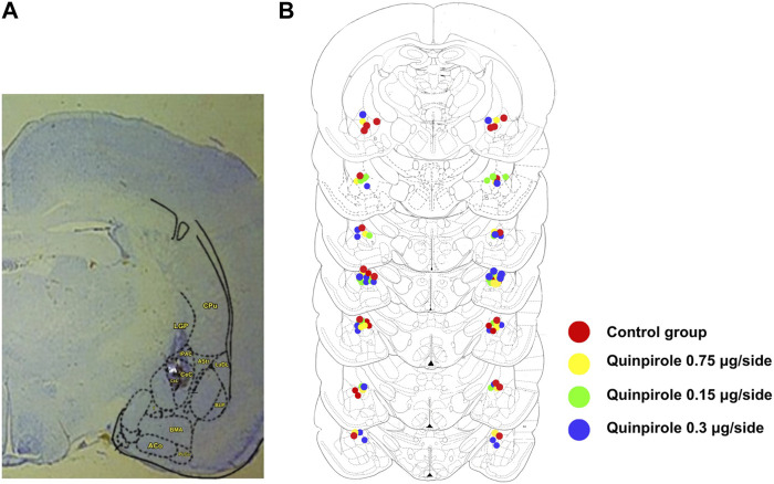
FIGURE 1.
Bilateral cannula placements within the amygdala. (A) Panel on the right shows a representative section within the amygdala (Br −2.30) depicting a representative bilateral cannula placement. The star indicates the position of the CeA within the amygdala. (B) Schematic representation of the sites of the bilateral cannula placements within the amygdala as verified by histological examination in rats infused with either saline or different doses of quinpirole (0.075–0.3 µg/side). Stereotaxic levels were taken from the rat brain atlas of Paxinos and Watson (1986). Overlapping of cannula placements has been produced in some sections due to the high density of injection sites. A similar distribution pattern of injection sites was observed in all other experiments carried out in this work.

