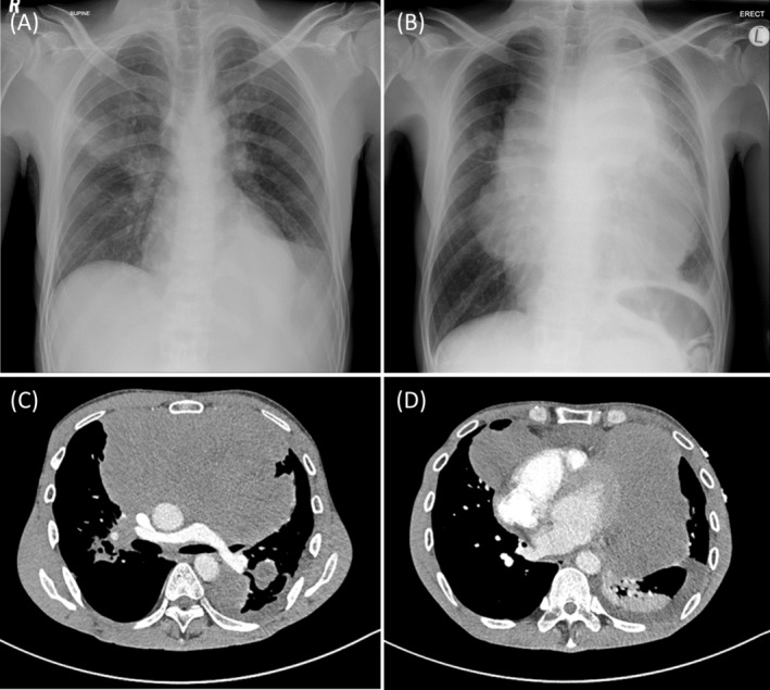
FIGURE 1.

(A) A supine AP chest radiograph taken 7 months prior did not show mediastinal widening. (B) An erect PA chest radiograph taken at this presentation demonstrated a very large, multi‐lobulated central mass causing significant mediastinal widening. An axial post‐contrasted CT chest at the subcarinal level (C) and at the level of the heart (D) showed this non‐enhancing mass to be centred in the anterior mediastinum and to displace and encase mediastinal structures including the pulmonary arteries, the aorta and the heart.
