FIGURE 2.

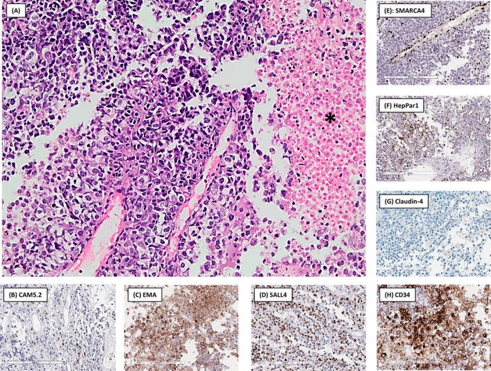
(A) The tumour was composed of sheets of epithelioid cells with clear cytoplasm. Tumour necrosis, a prominent finding in this specimen, is present on the right, indicated by a black asterisk (haematoxylin and eosin stain, original magnification 200×). Immunohistochemistry showed only focal positive staining in isolated cells with CAM5.2 (B), while convincing cytoplasmic expression was found with EMA (C) and nuclear expression with SALL4 (D). SMARCA4 (E) was completely lost in the tumour cells while still being expressed in endothelial cells (internal control). HepPar1 (F) showed patchy granular cytoplasmic staining in ±10% of tumour cells. Claudin‐4 (G) was completely lost while CD34 (H) stained positive in the majority of tumour cells.
