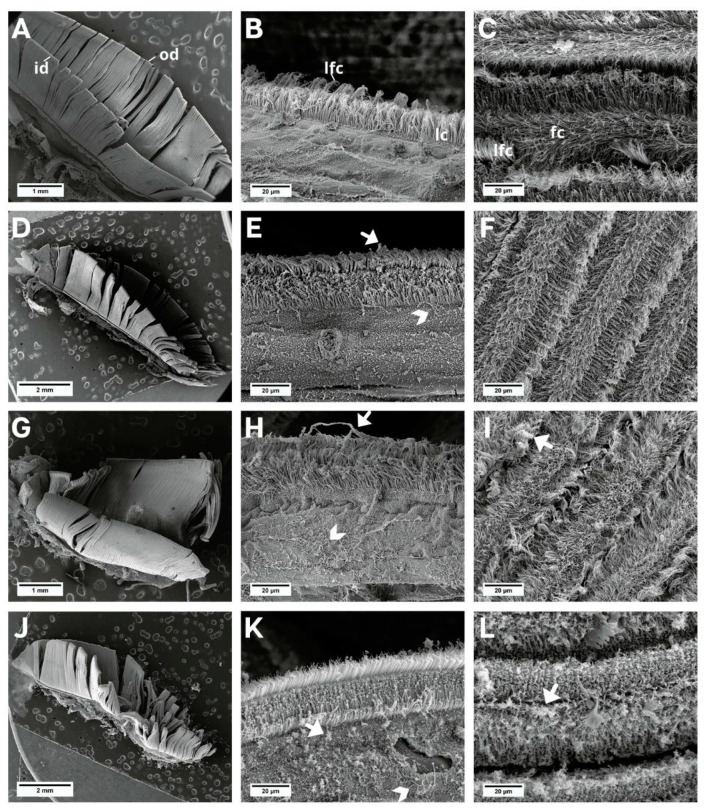
Figure 2.
SEM images of the L. fortunei gills. (A–C) Control group and (D–L) groups exposed to NaDCC. (A) Ciliated gill with inner (id) and outer demibranch (od). (B) Dorsal view of gill lamellae showing the frontal region of the filaments with lateral cilia (lc) and laterofrontal cirri (lfc). (C) View in perspective of a set of filaments, showing frontal cilia (fc) and laterofrontal cirri (lfc). (D–F) Group 24 h after conditional exposure. (G–I) Group 48 h after conditional exposure. (J–L) Group 72 h after conditional exposure (presumably they were dead, as their valves were open). Mucus (white arrow); loose cilia (arrow head).

