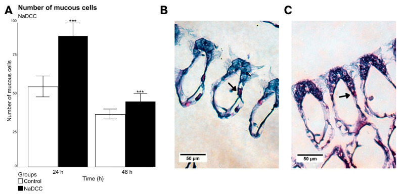
Figure 5.
Mean and standard deviation of the number of mucous cells present in 50 branchial filaments to NaDCC exposed (24 and 48 h after conditional exposure) (A). Significant differences between exposure groups are represented by *** p < 0.01. Group exposed for 72 h presented a mortality rate of almost 100%, as their valves were open. (B,C) Light micrograph images showing the transverse sections of a gill filament of L. fortunei stained with AB-PAS. (B) Group 24 h after conditional exposure. (C) Group 48 h after conditional exposure. Mucus-producing cells (arrow).

