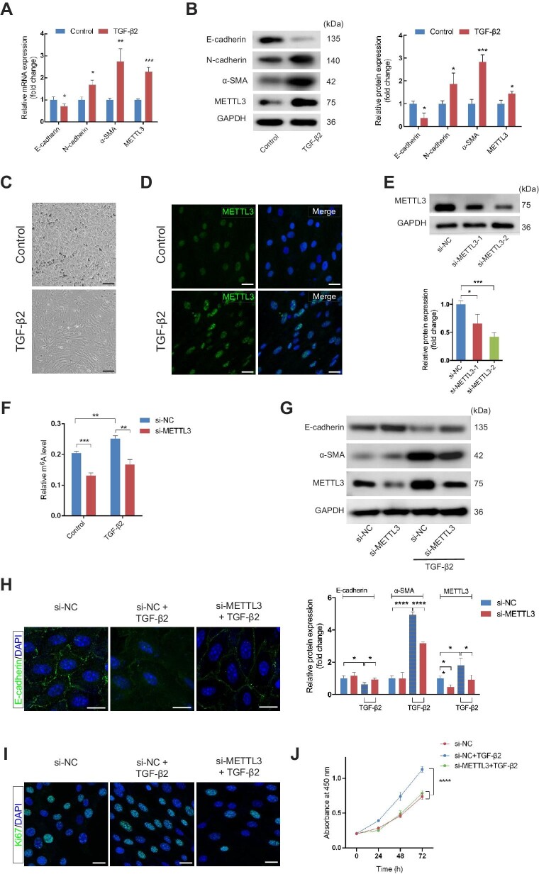
Figure 3.
METTL3 is involved in primary mouse RPE cells undergoing EMT. (A and B) The mRNA (A) and protein (B) levels of EMT-related markers (E-cadherin, N-cadherin, and α-SMA) and METTL3 in primary mouse RPE cells with and without 10 ng/ml TGF-β2 for 48 h. (C) Phase-contrast microscopy images of RPE cells showed the spindle fibroblast-like morphology induced by TGF-β2 application. Scale bar, 50 µm. (D) Immunofluorescence analysis demonstrated increased METTL3 in RPE cells treated with TGF-β2. Scale bar, 25 µm. (E) Primary RPE cells were transfected with si-NC, si-METTL3-1, or si-METTL3-2. METTL3 protein expression was quantified by western blotting after 48 h. (F) Primary RPE cells were transfected with si-NC or si-METTL3-2 for 48 h before the application of TGF-β2 or not. Then, total RNA was extracted to assess the global m6A level. (G) Western blotting analysis of E-cadherin and α-SMA was performed to determine the effect of METTL3 inhibition on TGF-β2-induced EMT in primary RPE cells. (H) Immunofluorescence analysis of E-cadherin in RPE cells confirmed that the loss of E-cadherin caused by TGF-β2-induced EMT was partially restored by METTL3 deficiency. Scale bar, 25 µm. (I) Ki67-positive cells were reduced after METTL3 inhibition. Scale bar, 25 µm. (J) CCK8 assay showed that cell proliferation was inhibited by METTL3 knockdown. Data present mean ± SD of three independent experiments. Student's t-test for two independent groups and two-way ANOVA tests for CCK8 assay, *P < 0.05, **P < 0.01, ***P < 0.001, ****P < 0.0001.

