Figure 1.
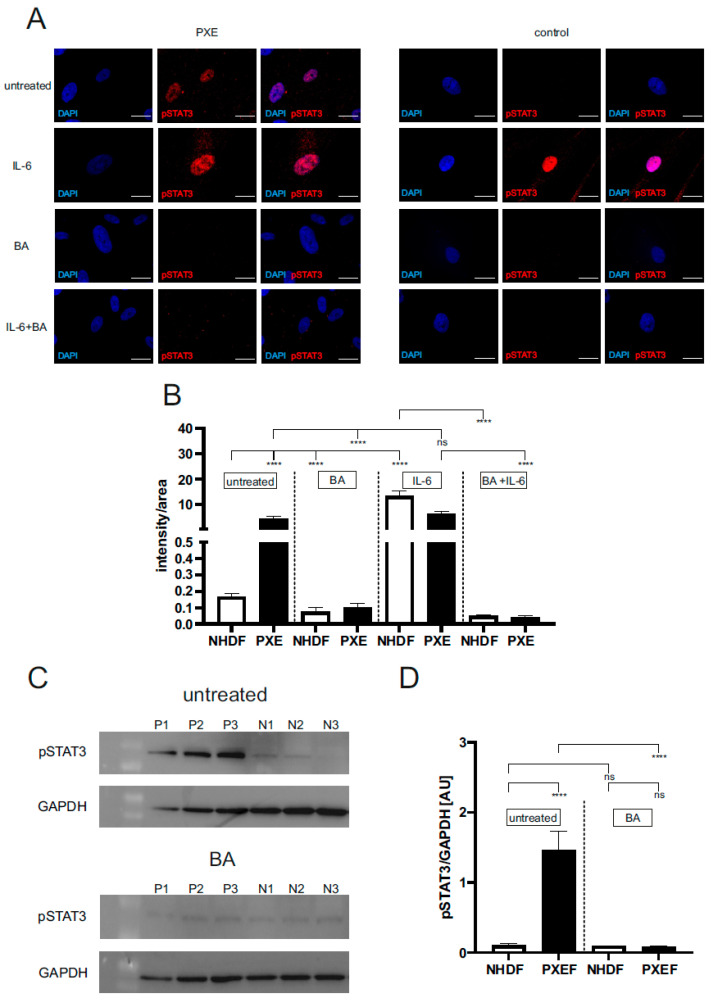
Immunofluorescence and Western blot of pSTAT3 (pTyr705). Dermal fibroblasts from patients with pseudoxanthoma elasticum (PXEF) (n = 4) and normal human dermal fibroblasts (NHDF) (n = 4) were cultivated for 72 h in medium with 10% lipoprotein deficient fetal calf serum (LPDS). Fibroblasts were treated only with DMSO (untreated), 1 µM baricitinib (BA), 50 ng/mL interleukin-6 (IL-6) or together (BA + IL-6). (A) Immunofluorescence analysis of pSTAT3 (red); cells were counterstained with DAPI (blue). (B) Quantification of pSTAT3 (intensity/area) in PXEF (black) and NHDF (white). (C) Western blot analysis of pSTAT3 expression of PXE (P) and NHDF (N). (D) Quantification of pSTAT3 normalized on GAPDH expression shown in arbitrary unit (AU) in PXE (black) and control (white) fibroblasts. Representative images are shown at 100× magnification (A, scale bar 15 µm). Data are shown as mean ± SEM. **** p ≤ 0.0001, ns p > 0.05.

