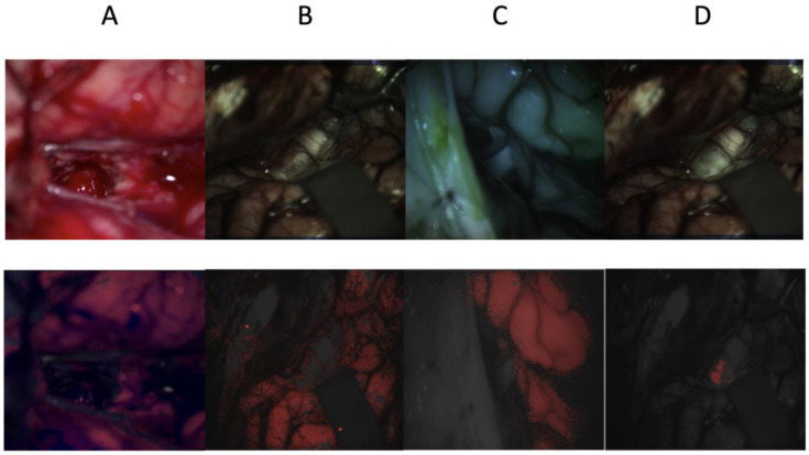
Figure 5.
Pediatric brain images were captured using RGB, visible, and infrared HSI cameras. The four images at the top are the original RGB and hyperspectral images that have been artificially colored. The bottom four images are their respective segmentations overlayed in red. Images (A–C) are collected from the RGB, visible HSI, and infrared HSI datasets, respectively, where the healthy tissue is being segmented, and image (D) is obtained from the visible HSI dataset but for tumor segmentation.

