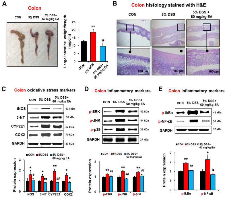
Figure 4.
EA treatment reduced oxidative/nitrative stress and NF-κB/MAPK signals in the colon of DSS-induced IBD mice. (A) Representative of macroscopic images and length of large intestines, as indicated. (B) Representative H/E-stained images of formalin-fixed colon sections in the indicated groups. (C) The levels of colon iNOS, nitrated proteins detected by anti-3-NT antibodies, CYP2E1, and COX2 in the indicated groups are presented. (D,E) The levels of p-ERK, p-JNK, p-p38, p-IκBα, and p-NF-κBp65 in the indicated groups are presented. Data represent means ± S.E.M. (n = 5–7/group). The statistical significance between values for each group was assessed by Dunnett’s t-test. * p < 0.05, ** p < 0.01 between 5% DSS and control groups; # p < 0.05, ## p < 0.01 between 5% DSS vs. 60 mg/kg EA groups.

