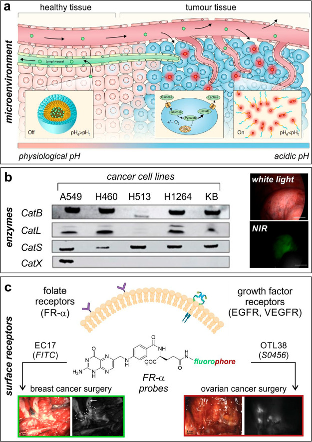
Figure 2.
Biological targets for translational fluorescence imaging in humans. (a) Extracellular cancer acidosis as a generic target for a broad range of solid tumors.41 The tumor microenvironment turns acidic when the cancerous tissue becomes invasive; this is exploited for the ONM100 nanoparticles to extravasate because of enhanced permeability of the tumor vasculature and results in ONM-100 accumulation within the acidic extracellular matrix with a concomitant switch from the “off” (green) to the “on” (red) state. Reproduced under a Creative Commons license from ref (41). (b) Cathepsin-activatable agents for in-human imaging of cancer cells.24 Western blot analysis of cathepsin expression in human nonsmall lung cancer cells using KB human cervical carcinoma cells as a positive control. Representative white light (top) and NIR fluorescence (bottom) images of a pulmonary tumor after administration of the cathepsin-activatable VGT-309 probe. Scale bars: 1 cm. Reproduced from ref (24) with permission from the American Association for Cancer Research. (c) FR-α and growth factor receptors as potential biological targets for imaging of cancer tissue. (Left) FR-α probe EC17 (fluorophore: FITC) was employed during breast cancer surgery to identify a bisected primary breast cancer lesion by using fluorescence imaging (dashed circles). The white arrow indicates tissue autofluorescence signals.48 Reproduced under a Creative Commons license from ref (48). (Right) the FR-α probe OTL38 (fluorophore: S0456) was employed during ovarian cancer surgery to identify retroperitoneal lymph nodes containing metastases of ovarian cancer.49 Reproduced from ref (49) with permission from the American Association for Cancer Research.

