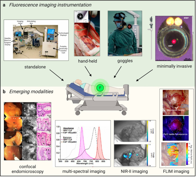
Figure 3.
Maximizing the clinical impact of fluorophores. (a) Fluorescence imaging instrumentation used in the clinic. (Left to right) The FLARE imaging system deployed in the operating room.59 Reproduced from ref (59) with permission from Springer Nature. Hand-held device placed on a measurement site in a brain to detect PpIX fluorescence in patients with suspected glioblastoma.65 Reproduced from ref (65) with permission from Elsevier. Fluorescence goggles worn by a surgeon to image ICG in patients during hepatocellular carcinoma (HCC) resection.67 Reproduced from ref (67) with permission from Elsevier. Minimally invasive procedures with a graphical representation of a laparoscope and an en face view of a flexible NIR cholangioscope made of a single-mode fiber that delivers laser excitation and 2 multimode fibers (MMFs) that collect light.72 Reproduced from ref (72) with permission from Elsevier. (b) Emerging imaging modalities. (Left to right) Confocal endomicroscopy images of colonic mucosa in patients with IBD.74 Reproduced under a Creative Commons license from ref (74). Representative white light images (left panels), confocal endomicroscopy (middle panels), and histology images (right panels) are shown in decreasing order for adenoma, hyperplastic polyps, ulcerative colitis, and Crohn’s disease. Multispectral imaging featuring absorbance and emission spectra of the fluorescent peptides QRH*-Cy5 and KSP*-IRDye800 for multiplexed imaging in Barrett’s esophagus patients.76 Reproduced from ref (76) with permission from BMJ Publishing Group Ltd. NIR-II imaging of ICG in a HCC tumor after resection highlights no remaining signal left in the NIR-I window (top panel) but remaining malignant tissue in the NIR-II window (bottom panel).77 Reproduced from ref (77) with permission from Springer Nature. FLIM imaging of a superficial glioblastoma tumor using PpIX78 with white light image of the surgical field of view (top panel), standard fluorescence microscope image used for 5-ALA visualization (excitation 405 nm) (middle panel), and fluorescence lifetime image of the PpIX channel (629 nm/653 nm, bottom panel). Reproduced under a Creative Commons license from ref (78).

