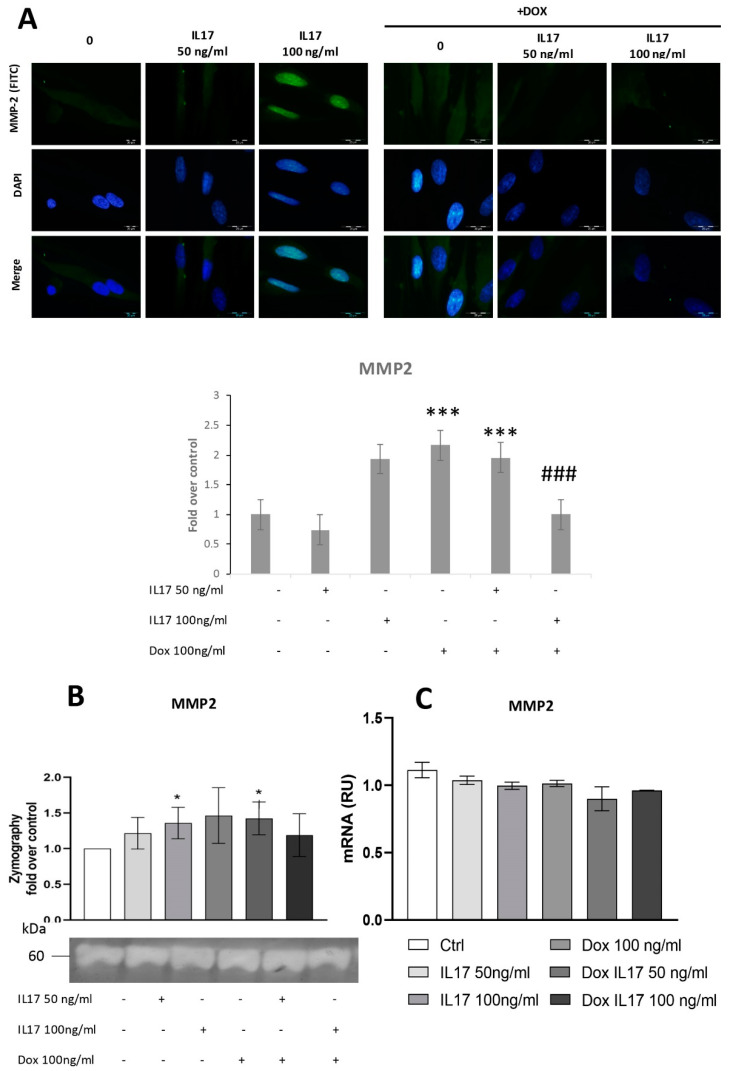
Figure 3.
Dox inhibits MMP2 expression of PDLSCs, abrogating the effect of IL-17. The cells were treated for 24 h with 0, 50 and 100 ng/mL IL-17 with or without Dox 100 ng/mL: (A) Expression of MMP2 in PDLSCs analyzed by immunostaining with MMP2 primary antibody, corresponding FITC-conjugated secondary antibody and DAPI. Representative images of immunofluorescence microscopy are shown. Scale bars: 20 μm. Graphical: of MMP2 protein expression as analyzed by Image J, relative protein expression normalized to control. Results in graphs are presented as mean ± SEM from at least three independent experiments. Statistically significant differences: *** p < 0.001 compared to control, ### p < 0.001 compared to Dox. (B) MMP2 activity and protein expression were determined by zymography. Statistically significant differences: * p < 0.05 compared to control (C) MMP2 normalized to the Ct value of the housekeeping gene GAPDH. Results were presented as the mean ± SEM from at least two independent experiments.

