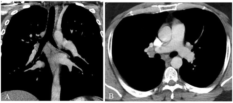
Figure 2.
Computed tomography of the thorax with contrast (soft tissue window settings) in coronal (A) and axial (B) planes. No nodules or areas of opacification were visualized within the lung parenchyma. Nonspecific subcarinal and right hilar lymphadenopathy was visualized in addition to prominent however nonenlarged left hilar lymph nodes.

