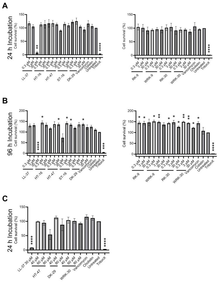
Figure 5.
Cytotoxicity of modified CLEC3A-derived peptides. (A) Cytotoxicity assay of NIH3T3 cells with LL-37 (30 µM ** p = 0.003), HT-16, HT-47, ST-16, DK-29, RK-9, WRK-9, RK-30, WRK-30, Vancomycin, Colistin, Untreated, and TritonX (**** p < 0.0001). (B) Cytotoxicity assay of NIH3T3 cells LL-37 (30 µM **** p < 0.0001), HT-16 (0.3 µM * p = 0.050), HT-47 (3 µM * p = 0.044), ST-16 (0.3 µM * p = 0.032, 3 µM * p = 0.040), DK-29 (3 µM * p = 0.025), RK-9 (0.3 µM * p = 0.038, 3 µM * p = 0.045), WRK-9 (0.3 µM * p = 0.016, 3 µM ** p = 0.010), RK-30 (0.3 µM * p = 0.011, 3 µM * p = 0.046, 30 µM * p = 0.035), WRK-30 (0.3 µM ** p = 0.005, 3 µM ** p = 0.006, 30 µM * p = 0.024), Vancomycin (* p = 0.035), Cholistin, Untreated, and TritonX (*** p = 0.0002, **** p < 0.0001). (C) Cytotoxicity assay of NIH3T3 cells with LL-37 (**** p < 0.0001), HT-47, DK-29, and WRK-30, Vancomycin, Colistin, Untreated, and TritonX (**** p < 0.0001). All values are percentages normalized to the untreated controls (untreated controls were set as 100%). Depicted are averages and standard deviations. Statistical significance was calculated using Prism 9 with a paired ANOVA test followed by a Dunett test and multiple comparisons, comparing each treatment with the untreated control. All experiments were repeated three times (n = 3).

