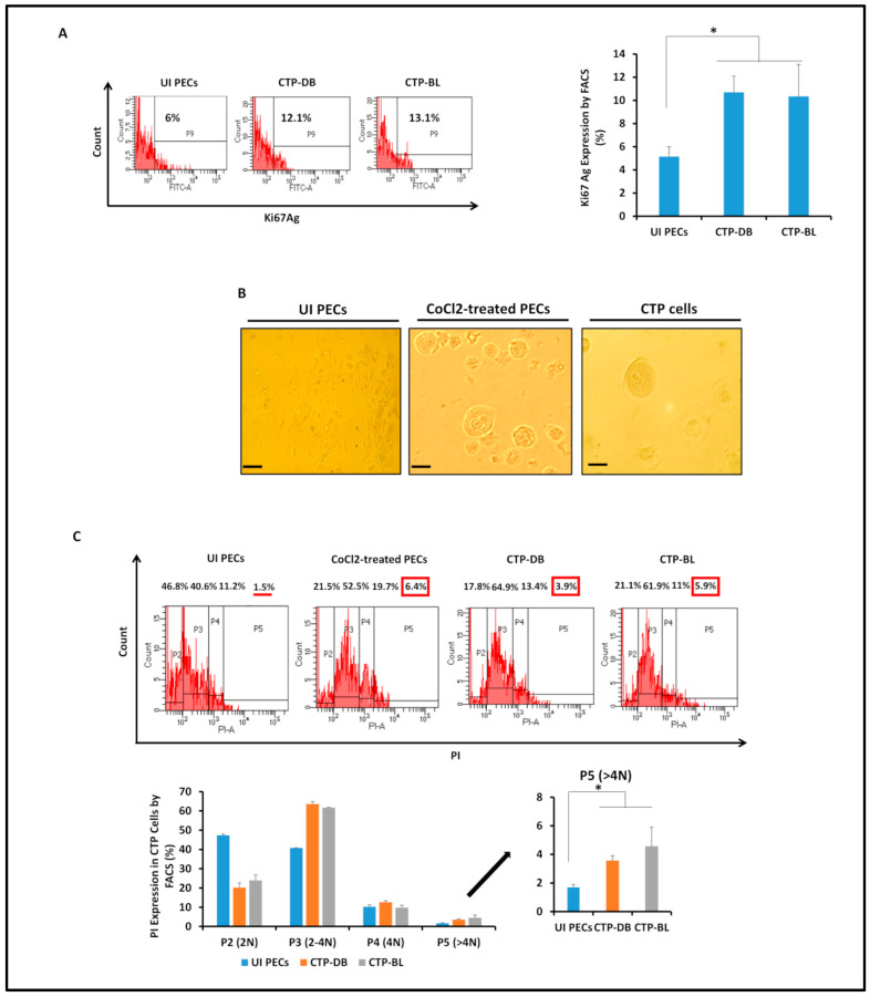
Figure 3.
Assessing the proliferative potential and detecting polyploidy in CTP-DB and -BL cultures. (A) FACS staining of Ki67Ag in uninfected PECs as well as CTP-DB and -BL cells. (B) Microscopic images of polyploidy detected in CTP-DB and -BL cultures. Cobalt chloride (CoCl2)-treated PECs (300 μM) were used as a positive control, while uninfected PECs were used as a negative control. Magnification ×200, scale bar 50 μm. (C) Propidium iodide (PI) staining for polyploidy detection in CTP-DB and -BL cells by FACS. CoCl2-treated PECs were used as a positive control, and uninfected PECs were used as a negative control. Data are represented as mean ± SD of two independent experiments. * p-value ≤ 0.05. The red line shows the low percentage of P5 (>4 N). Red boxes emphasizes the high percentages of p5 (>4 N).

